
Time-resolved serial crystallography on three GPCRs
From acting as starting points for virtual screening approaches, to providing information on ligand binding modes for lead development – experimentally obtained protein structures have been cemented as a crucial feature of the drug design process. However, despite their established use, protein structures still present limitations in terms of their relevance to the physiological system.
Time-resolved serial crystallography (TRSX) allows some of these limitations to be surmounted. In TRSX, structures are collected at room temperature, where differences in ligand binding, loop positioning, and side chain orientations relative to structures collected at cryogenic temperatures have previously been observed. Additionally, TRSX allows for the determination of high-resolution structures of transient intermediate states in reaction pathways. These previously unobserved states can help to enhance knowledge on dynamic processes or serve as better starting points for drug design.
Together with the Standfuss group at the Paul Scherrer Institute, time-resolved crystallography was carried out for three G protein-coupled receptors (GPCRs). GPCRs are pharmacologically relevant protein targets, with the three proteins studied: A2A, β2AR and mGlu5, being involved in Parkinson’s, lung and cardiovascular, and neurodegenerative diseases respectively.
To facilitate the study of these receptors, synthetic photoswitches were used to introduce a light-activated trigger. These photoswitches undergo light-induced isomerization from high to low affinity states, enabling the study of processes such as ligand dissociation.
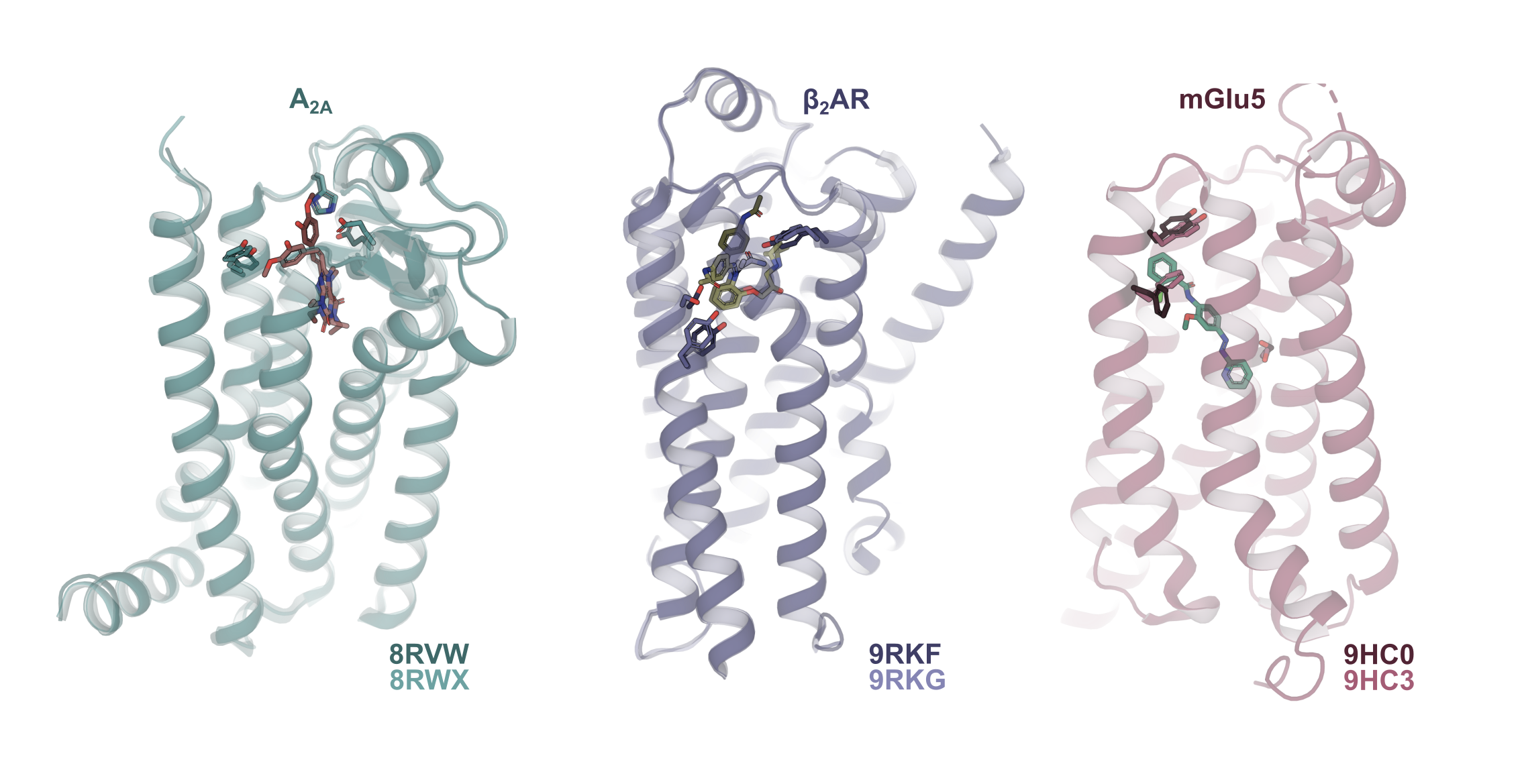
In the A2A receptor, steady state data were collected at the SLS using 3 different designed synthetic photoswitches. Differences in the chemical structures of the photoswitches affected their behavior in the receptor after isomerization, with the largest changes observed with StilSwitch3; including opening of the canonical binding pocket gate between Glu169 and His264 and movement of Tyr271 to accommodate the isomerized ligand in the binding pocket. Furthermore, the loss of cholesterol from the receptor, which binds specifically to the inactive state, indicates the propagation of changes throughout the receptor following ligand dissociation. A follow up experiment at MaxIV to collect time-resolved data suggested the existence of two states following illumination: the bound isomerized ligand at 2-20 ms, and the formation of the apo state at 50-70 ms. Work is currently ongoing in collaboration with the Standfuss group to improve the resolution of these data to enable the accurate modelling of these states.
For the β2AR, ligand isomerization was not followed by release from the binding pocket. Instead, the isomerized ligand adopts a new pose in the binding pocket, causing rearrangement of residues in the binding pocket and shifting the positions of helices 5, 6, and 7 towards a state reminiscent of agonist-bound structures of β-adrenergic receptors. Additionally, a key interaction between Asn293 and Ser204 is broken by the isomerized ligand in a way typical of agonist binding to the β2AR. Subsequent activity assays revealed that upon isomerization, the photoswitch is converted from an inverse agonist to a neutral antagonist.
For mGlu5, multiple timepoints were collected between 50 ms and 8 s using both annealing and the Spitrobot crystal plunger (developed by researchers from Hamburg) in combination with laser triggering. Using single crystals to collect time-resolved data, some of the challenges of TRSX could be avoided, namely the requirement for largescale production of crystals, allowing for the study of more challenging-to-produce proteins. Following illumination, loss of the ligand from the allosteric binding pocket of mGlu5 could be observed. This dissociation is coupled with rearrangements in the protein to adapt to the apo state of the protein, including the binding of waters and movements of sidechains to fill the empty binding pocket.
Together, these findings demonstrate the exciting opportunity offered by TRSX to study the structural dynamics of protein-ligand interaction to better understand induced fit structural changes and improve in silico ligand screening as well as design methods further.
