
leadXpro Therapeutics Portfolio






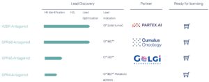
Selective Adenosine 2B Receptor Antagonists
Adenosine Receptors, a GPCR family with four subfamily members (A1, A2A, A2B, A3), are homeostatic sensors that respond to cellular stress, hypoxia and inflammation. The adenosine receptor 2B subtype (A2BR) is the only low-affinity (<1 mM) adenosine receptor. It remains silent during normal homeostasis and signals exclusively in situations that involve pathological high-adenosine levels.
In the Tumor Microenvironment, adenosine acts as a natural “off switch” for the immune system and the A2BR was identified as a key driver of this suppression. Preclinical data suggests that A2BR antagonism can boost the effectiveness of existing cancer immunotherapies.
In Multipe Sclerosis (MS), A2BR signaling promotes pathological Th17 cell differentiation, increases inflammatory IL-6 release, inhibits oligodendrocyte maturation and suppresses remyelinisation.
(see also: Partex.AI and leadXpro collaboration announcement)
Proton-sensing GPCRs, also called pH-sensing GPCRs, are a subgroup of G-protein-coupled receptors that respond to extracellular acidification and convert it into intracellular signals. Known members include GPR4, GPR65, GPR68 and GPR132.
Their pH sensitivity is mediated largely by protonatable extracellular amino acid residues, especially histidines, which change protonation state as extracellular pH falls. When the pH drops from physiological pH (~7.4) into the mildly acidic range (<7.4):
- extracellular residues become protonated
- the receptor undergoes conformational changes
- G-protein signaling is activated
Because tissue acidosis occurs across a wide range of pathological states, proton-sensing GPCRs are emerging as important regulators in inflammation, ischemia, tumor microenvironments, and tissue injury. They are also active in certain physiological settings, such as exercising skeletal muscle exercise, when local pH shifts drive adaptive responses.

GPR68
GPR68 is the most pH-sensitive member of the family, activated when tissue pH drops below pH7.3. Through control of cytokine release, fibrosis, and immune cell reprogramming, GPR68 drives the biological processes underpinning inflammatory and fibrotic processes in inflammatory bowel disease (IBD) and colorectal cancer.
Due to insufficient blood perfusion, hypoxia, inflammation, and glycolytic cell metabolism, the tumor microenvironment (TME) is characteristically acidic. The acidification of the TME, due to the increased production of lactate and protons, promotes chemoresistance and drives immune evasion and cancer progression. GPR68 is upregulated in both breast cancer and head and neck cancer when compared to control tissue and in both cases increased GPR68 correlates with worse outcome. Moreover, GPR68 inhibition reduces tumor growth in preclinical models, highlighting GPR68 as a promising therapeutic target in oncology. (source: Cornell J, et al. J Cancer Biol. 2024;5(2):65-75)
GPR68 expression is substantially upregulated in the inflamed intestinal mucosa of patients with IBD. GPR68 gene knockout or pharmacological inhibition of GPR68 has been shown to ameliorate colitis in preclinical models, highlighting GPR68 as a promising therapeutic target in IBD. (source: Hausmann M, et al. Pflugers Arch. 2024;476(4):611-622)

GPR65
GPR65 is a proton-sensing Gas-coupled receptor predominantly expressed in immune cells, including T cells, macrophages, NK cells, and neutrophils. It functions as a pH-dependent regulator of immune cell activity and becomes activated under mildly acidic conditions (pH ~7.0–6.5).
GPR65 activation raises intracellular cAMP, which suppresses downstream TCR signalling and dampens pro-inflammatory cytokine production (TNF-α, IL-6). In physiological acidosis – such as in inflamed or ischaemic tissue – this may function as a homeostatic brake on excessive inflammation. In the tumour microenvironment, however, this same mechanism is co-opted by tumour acidity to suppress cytotoxic T-cell and NK-cell activity, contributing to immune evasion. GPR65 was associated with poorer overall survival and primary treatment outcome in patients with lung adenocarcinoma. GPR65 expression was found to be closely correlated with multiple tumor infiltrating immune cells (TIICs) and immune checkpoint molecules. GPR65 antagonism has been shown to restore antigen-specific T-cell killing in vitro without direct cytotoxicity on tumour cells. (source: Zhou H, et al. Front. Immunol. 2025; 16:1572757)

GPR4
GPR4 is a Gs-coupled proton-sensing receptor that converts extracellular acidosis into intracellular cAMP signalling. It is most highly expressed in vascular endothelial cells, with notable expression in lung, kidney, and the retrotrapezoid nucleus (RTN) of the brainstem.
In the tumour microenvironment, persistent acidosis chronically activates GPR4 on endothelial cells, promoting pathological angiogenesis. GPR4 activation also impaired CD8+ T cell infiltration into tumor nests and fostered immune exclusion in the tumor microenvironment in colon tumor models through the JAK2/STAT3 signaling pathway. (source: Bai, S. et al. Nat. Commun. 2026;17: 1196)
In the inflamed intestinal mucosa, GPR4 upregulation correlates with colitis severity. In preclinical knockout models, GPR4 deficiency reduced intestinal inflammation and limited colitis-associated colorectal cancer development. (source: Sanderlin, E.J. Biochim. Biophys. Acta. 2017;1863(2):569-584)
The augmented utilization of lipids for energy supply is accelerated in the liver, skeletal muscles, and adipose tissues with insulin resistance. This process leads to high production of protons, frequently observed in diabetic patients. GPR4 activation upregulates leukocyte recruitment and inflammation. GPR4 antagonists could help to attenuate chronic vascular inflammation in patients with metabolic disease. (source: Dong L, PLoS One. 2013;8(4):e61991)
